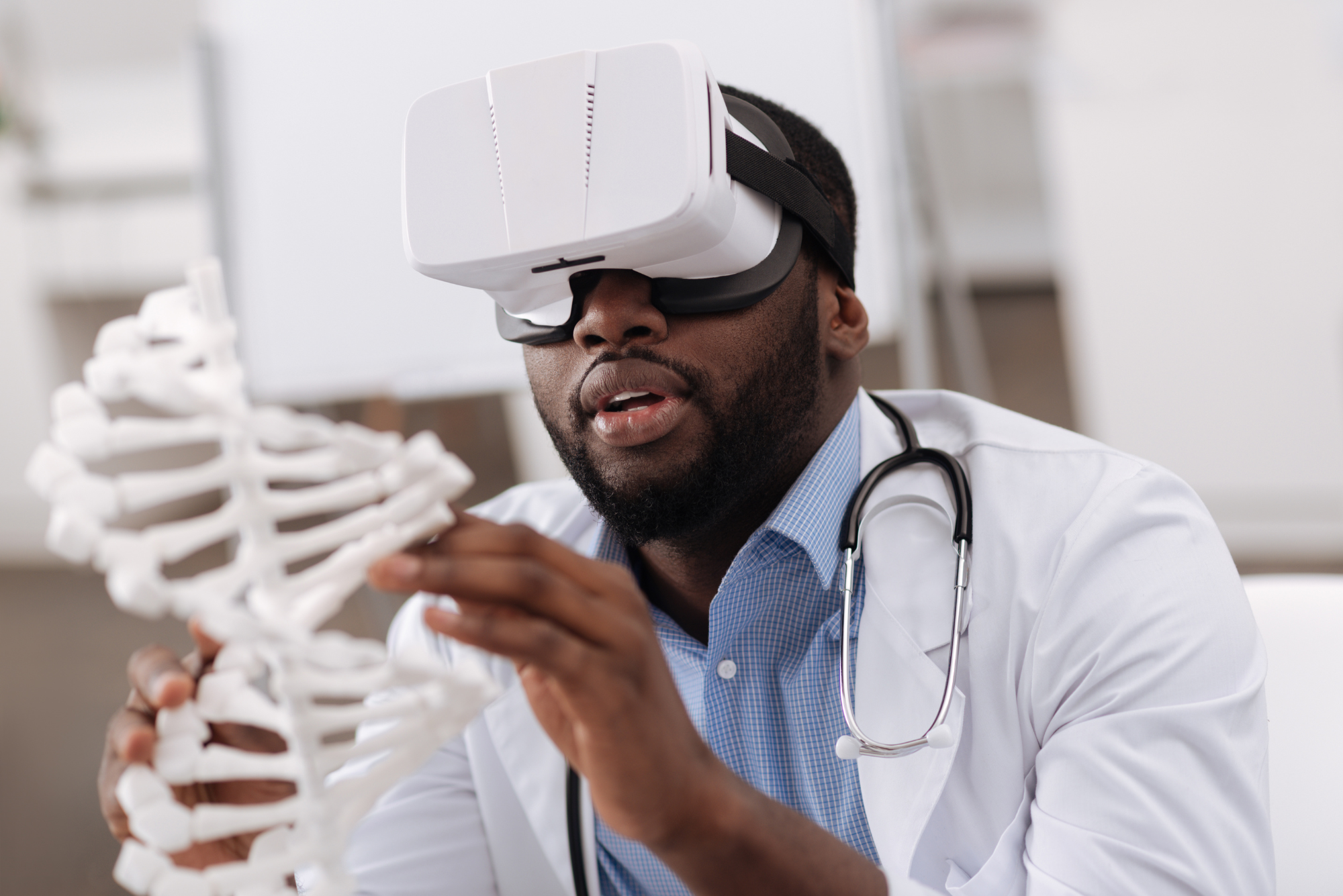
Like many doctors fascinated by new medical technology, I have succumbed to “me-search”: the urge to use new technology to study myself. Me-search optimizes my own health and teaches me how to use new technology to help patients.
Last October at a medical conference, I learned about Empower Sleep, a new sleep medicine telehealth company. Empower Sleep promises to transform sleep medicine using a device called the SleepImage Ring.
I’ve always struggled with sleep due to a smorgasbord of areas for improvement:
- Nasal obstruction due to hay fever
- Bruxism (nocturnal teeth grinding)
- Nocturia (arising more than once a night to urinate)
- Light sleeping (more sensitive to disruption from environmental stimuli)
- Advanced sleep phase disorder (arising too early in the morning)
- Preterminal insomnia (awakening before the end of the night, often associated with depressive illness)
Me-search overtook me. I had to try the SleepImage Ring for myself.
(Disclaimer: I have no financial interest in SleepImage or Empower Sleep. I do refer patients to Empower Sleep for treatment.)
What We’ve Learned About Sleep
From time immemorial, sleep was thought to be an inactive state between wakefulness and death. Sleep was understood as a passive, inactive state defined by lack of consciousness. Dreams represented a peek into this realm and were thought to carry mystical, psychological, and/or religious meaning.
In 1928, we gained the ability to peer into sleep’s biology when German psychiatrist Hans Berger developed a way to record the electrical activity of the brain’s neurons. The electroencephalogram (EEG) revealed distinctive waves of neuronal activity unique to sleep.
By the 1950s, an EEG-defined sleep phase associated with rapid eye movements (REM) and dreaming had been characterized, and the concept of sleep architecture was born. By the 1960s, EEG-based polysomnograms (PSG) were used to diagnose and treat sleep disorders.
We now know that sleep occurs in 90-minute cycles that repeat through the night. Each cycle contains lighter portions, S1 and S2, and deeper portions, S3, called slow-wave sleep (SWS). Each cycle also contains REM sleep and brief arousals.
The composition of those 90-minute cycles changes throughout the night: more slow-wave sleep occurs early, and more REM sleep occurs later. The amount of slow-wave sleep diminishes as we age, while the percentage of REM sleep is preserved.
Many other factors can affect sleep’s composition, including:
- Illnesses such as obstructive sleep apnea (OSA)
- Medications such as benzodiazepines (drugs like Valium)
- Beverages such as alcohol
The Current State of Sleep Testing
EEG-based polysomnograms have been the dominant method of diagnosing sleep disorders for over 50 years, forming the foundation of sleep medicine. However, a polysomnogram is an extremely cumbersome test.

The patient must travel to a special overnight clinic and fall asleep with 22 separate wires attached to their body, which measure:
- Brain wave activity
- Airflow
- Eye movement
- Oxygen saturation
- Heart rate and rhythm
- Muscle tone in the chin, chest wall, and legs
Newer home sleep studies have reduced the number of attachments needed and allowed the patient to sleep in their own bed. However, both in-clinic and at-home studies may influence the sleep being measured.
Think about how well you slept last week. For most of us, the night-to-night experience of sleep varies. Any single night of sleep (let alone with 22 wires attached!) may not represent a typical night.
Neither in-clinic nor at-home tests are practical for multiple nights of testing. Repeating a study allows for the assessment of treatment modalities and sets up a series of trial-and-error experiments to optimize sleep.
Fortunately, we’re amidst a quiet revolution in our understanding of sleep’s physiology.
The Sleep Revolution
For the last hundred years, we’ve classified sleep based on the brain’s electrical activity as measured by an electroencephalogram. However, sleep entails not just brain activity but characteristic changes throughout the entire body, including breathing, heart rate, blood pressure, and body temperature.
About 15 years ago, researchers began developing a new method for analyzing sleep based on changes that occur to our autonomic nervous system (ANS). The autonomic nervous system is a network of nerves that regulate unconscious processes that keep our body functioning, including breathing, digestion, and heart pumping.
The autonomic nervous system is divided into two categories: the sympathetic (SNS) and parasympathetic (PNS) nervous systems:
- The sympathetic nervous system mediates our “fight or flight” response — increasing our breathing rate, shunting blood to our muscles, making the heart pump harder, and putting more fuel into the bloodstream for muscular activity.
- The parasympathetic nervous system mediates “rest and digest” functions such as improving blood flow to the digestive tract and secreting digestive enzymes.
The sympathetic and parasympathetic nervous systems exist in a delicate balance, as every organ receives signals from both systems.
For example, in the cardiopulmonary system (heart and lungs), signals oscillate back and forth in a teeter-totter fashion, and each breath subtly alters the heart rate:
- During each breath in, called inspiration, the sympathetic nervous system predominates and slightly speeds up the heartbeat.
- During each breath out, called expiration, the parasympathetic system slightly predominates and slightly slows down the heartbeat.
Thus, continuous recording of every heartbeat demonstrates variation in timing that correlates with the respiration rate. Exactly how much variability reflects the strength of the sympathetic nervous system’s input relative to the parasympathetic nervous system’s. A stronger parasympathetic nervous system creates more variability and correlates with better health outcomes.
Breathing also affects the amount of blood pumped each time the heart contracts. Each heartbeat during expiration moves a bit more blood than each heartbeat during inspiration.
A new technique called cardiopulmonary coupling (CPC) generates one waveform reflecting the change in heart rate and a second waveform reflecting the change in blood output. By analyzing the relationship between these two waveforms, researchers assess how the autonomic nervous system functions during sleep.
Cardiopulmonary coupling is a brand-new lens we can use to view sleep. EEG describes sleep from the vantage point of the brain’s wave changes, while cardiopulmonary coupling divides sleep into different stages based on differences in the autonomic nervous system.
CPC vs. PSG: What’s the Difference?
Cardiopulmonary coupling results and polysomnogram results classify non-REM sleep (NREM) differently.
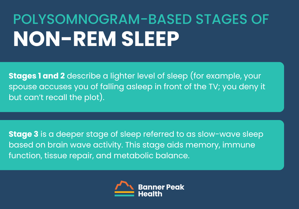
The traditional polysomnogram-based classification divides non-REM sleep into three stages of progressive depth:
- Stages 1 and 2 describe a lighter level of sleep (for example, your spouse accuses you of falling asleep in front of the TV; you deny it but can’t recall the plot).
- Stage 3 is a deeper stage of sleep referred to as slow-wave sleep based on brain wave activity. This stage aids memory, immune function, tissue repair, and metabolic balance.
Meanwhile, cardiopulmonary coupling categorizes non-REM sleep into stable and non-stable components. Stable non-REM correlates with slow-wave sleep but possibly provides a more accurate measure of this physiologically vital sleep stage.
The biggest advantage of cardiopulmonary coupling over polysomnograms is the ease of testing. A small rubber ring worn on your finger (like the SleepImage Ring, which uses cardiopulmonary coupling) provides almost as much information as 22 bulky wires (as in the case of a polysomnogram).
How Does the SleepImage Ring Work?
The SleepImage Ring harnesses an amazing new technology called photoplethysmography (PPG). Photoplethysmography powers our current array of consumer wearable devices, including the Fitbit, Garmin watch, Oura Ring, and WHOOP band, as well as medical grade, FDA-approved wearables such as the SleepImage Ring.
Photoplethysmography shines very small LED lights into the skin, illuminating tiny arteries under the surface. Adjacent sensors measure the reflected LED light, tracking the pulsatile pressure waves in the subcutaneous arteries and the color of the hemoglobin (oxygen-carrying protein) in the blood.
The pressure waves’ timing and amplitude measure the autonomic nervous system’s performance, and the hemoglobin’s color in the blood measures how well the lungs deliver oxygen to the body. The SleepImage Ring can also measure nocturnal movement and skin temperature.
Combining these data points paints a thorough picture of a person’s sleep. How many awakenings were there overnight? How much time was spent in vital stages such as REM and stable non-REM? Are there episodes of low oxygen indicating obstructive sleep apnea?
My SleepImage Experience
I started using the SleepImage Ring, hoping to gain insights that would minimize my sleep challenges. To my dismay, I discovered two new areas for improvement I’d been unaware of: a mild case of obstructive sleep apnea and an imbalance of my autonomic nervous system during non-REM sleep.
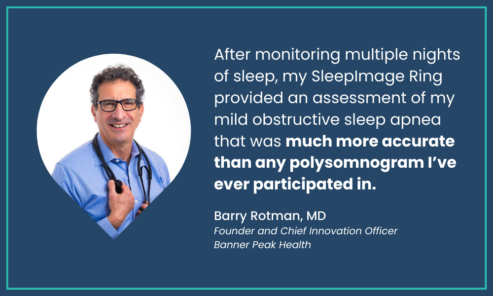
After monitoring multiple nights of sleep, my SleepImage Ring provided an assessment of my mild obstructive sleep apnea that was much more accurate than my one prior polysomnogram.
The SleepImage Ring tracked a tremendous amount of variation in the apnea-hypopnea index (AHI), the number of times per hour my breathing was compromised, ranging from 3 (normal) to 22 (moderate severity). My average was about 10, or mild.
Interestingly, in the traditional polysomnogram I underwent about 10 years ago, I struggled to sleep and the test revealed an apnea-hypopnea index of 5 (high normal). I suspect this is because the polysomnogram yielded only one night of compromised data.
Disheartened about receiving yet another sleep impairment diagnosis, I worked with Empower Sleep to use their cardiopulmonary coupling technology to address the problem. Not only is the SleepImage Ring better at diagnosing illnesses than polysomnograms are, but it also provides a better route for treating those illnesses.
Because my obstructive sleep apnea was mild, there was a good chance it could be treated without a CPAP machine. I was given a list of alternative therapies to try to reduce obstruction of my upper airway.
I incrementally added treatment modalities: nasal steroids to shrink the nasal mucosa and widen the opening of the nasal passages; lip tape to close my mouth, which pinched the tongue to prevent it from falling backward and obligated nasal breathing; and a soft rubber nasal device that further opens my nasal passages and reduces obstruction.
My bedtime routine has become more involved, and I’ve become quite a sight to behold in bed. But the process works! I now have several months of SleepImage data demonstrating the progressive reduction in my apnea-hypopnea index.
There’s still a fair amount of variation in my apnea-hypopnea index, but the average is now only a bit above normal. More importantly, I have more energy during the day.
My second discovery, an autonomic nervous system imbalance during non-REM sleep, represents a more complicated challenge because it’s a new disorder. Before the advent of cardiopulmonary coupling, autonomic nervous system impairments weren’t diagnosed by tracking sleep stages.
Because the disorder is so new, we don’t have good assessments of normal values associated with aging. There are only clues. We do know that EEG-based slow-wave sleep does somewhat correlate with the stable non-REM sleep stage.
We also know that slow-wave sleep declines with age. Therefore, we expect our autonomic nervous system’s function to decline, as well. Unfortunately, just as our skin gets “old and wrinkly” with age, so does our sleep.
Does my impaired autonomic nervous system function during sleep represent a normal age-related change, or can I improve it? As an athlete, I want to maximize the stable non-REM fraction of my sleep. That’s when “all the good stuff happens,” particularly the release of testosterone and growth hormone.
Unlike my clear victory over obstructive sleep apnea, I’m still working to overcome my impaired autonomic nervous system. I’ve expanded my meditation practice, targeting 20 minutes in the morning and 12 minutes before bedtime. (If you’d like to join me, review my step-by-step meditation guide.)
I’m also trying a very low dose of the antidepressant Lexapro, which I’ve used successfully to treat mild depression in the past. I’m not currently depressed, but there is a high probability that depression may be associated with a distinctive form of autonomic nervous system dysfunction during sleep.
My sleep data may represent a low-grade depression that medication can improve. The jury is still out on these interventions — I’ll keep you posted.
My Take on the SleepImage Ring
The SleepImage Ring represents a transformation in sleep medicine. More patients can be conveniently screened for sleep disorders, and more people can be effectively treated, especially those with obstructive sleep apnea.
Previously, only those with obvious obstructive sleep apnea symptoms underwent testing. Many people have the illness and don’t know it — I was one of them. They can now be diagnosed through greater use of sleep testing.
Also, less than half of people who are prescribed a CPAP machine to treat their obstructive sleep apnea can tolerate using it. Now we can use a trial-and-error method to build effective treatment regimens for those with mild and moderate obstructive sleep apnea, sparing them a CPAP machine.
The sleep medicine revolution has begun!